


The diagnosis of prostatitis includes more than 5 mandatory procedures and 4 additional procedures. Only a rectal examination of the prostate gland or ultrasound cannot tell for sure whether men have inflammation in the prostate. The reason is that many urological diseases have a similar clinical picture and only a comprehensive differential study eliminates the possibility of a wrong diagnosis.
How to pass an inspection
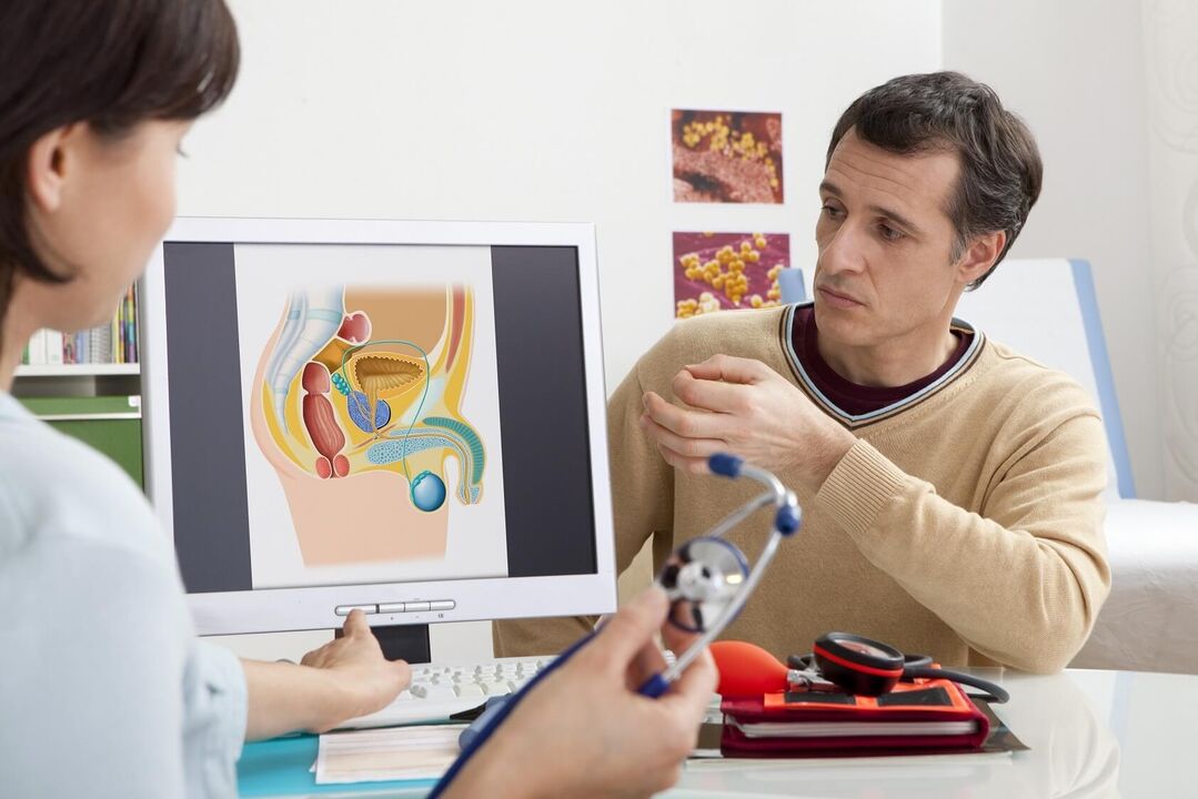
Men are recommended to have a preventive prostate examination by a urologist 1-2 times a year (prostatitis, adenoma and other prostate pathologies are asymptomatic in the early stages). When signs of the disease appear, you should immediately go to a specialist. Such symptoms are pain in the lower abdomen and groin, difficulty urinating and erection.
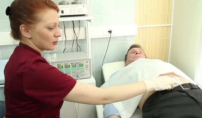
The doctor begins by collecting the patient's complaints and anamnesis, then performs a general examination. The next step in suspected prostatitis is a rectal examination (palpation of the prostate through a man's rectum). Finger research allows the doctor to assess the following parameters:
- Prostate size.
- Surface (smooth or bumpy).
- Density of the gland (soft or stony).
- The presence or tenderness of the central groove.
- A man's sensitivity when probing the prostate (if he experiences pain).
analyzes
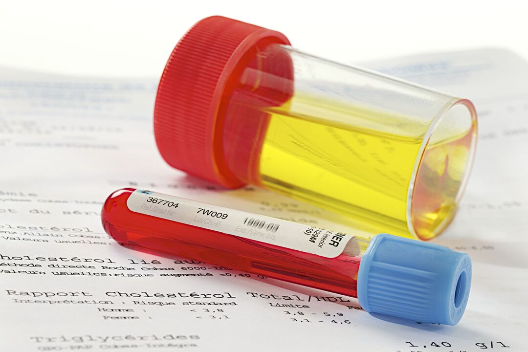
If a rectal examination and history taking suggest prostatitis, then the urologist's next step is to refer the patient for laboratory tests. According to clinical standards, the following types of examinations are mandatory:
- clinical analysis of urine;
- general blood analysis;
- urine planting for flora;
- when an infection is detected, the sensitivity of pathogens to antibiotics is determined.
A complete blood count helps to confirm acute prostatitis - with this diagnosis, there is an increase in the number of neutrophils with a shift of the leukocyte formula to the left and a strong decrease in the level of eosinophils. It is also possible to increase the ESR. Chronic inflammation is characterized by a low hemoglobin content (below 100 grams per liter of blood).
To rule out prostate cancer, a blood serum test is performed for the content of PSA - a prostate-specific antigen. Its increased amount indicates the presence of tumors, but does not determine their nature (benign or malignant). To detect this parameter, a prostate biopsy is performed with a histological study of the obtained material.
prostate secretion
During the rectal examination of the prostate, the urologist pays attention to the secreted secretion. Normally, it is thick, odorless, white in color. The maximum volume is 1-2 drops (3-5 ml). It should not contain impurities of pus or blood, as this is a sign of disease. The consistency of the liquid plays a role - if it comes out in clots, then the man has diverticular prostatitis. A more detailed study of the material allows laboratory research.
Microscopy and bacteriological study of prostate secretion is based on counting leukocytes, lecithin grains, amyloid bodies, macrophages, pathogenic and opportunistic organisms. Prostatitis is characterized by deviations:
- Acute prostatitis: the color of the secretion is yellow, the smell is sweet, the pH is acidic, there are less than half of the leukocytes and up to ¼ of the epithelial cells.
- Chronic bacterial prostatitis: yellow or brown color, sour smell, sour pH, less than half of leukocytes, macrophages (over 15), many amyloid bodies.
- Chronic non-bacterial prostatitis: the color is reddish, brown, there is no smell, leukocytes are normal, macrophages (10-20) are detected, there are many amyloid bodies.
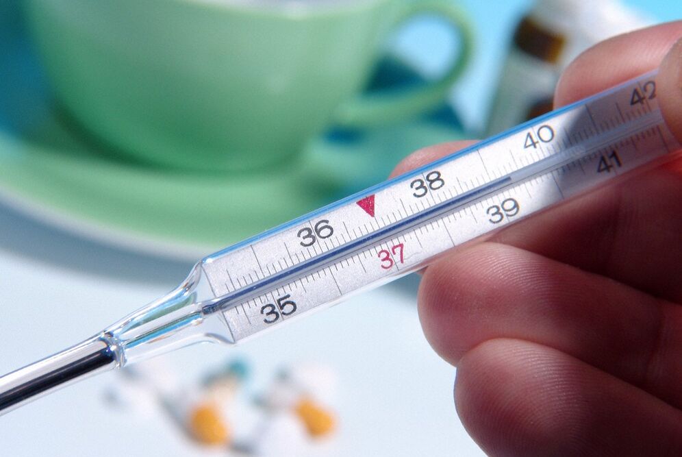
In some cases, the study of the secretion does not allow the detection of prostatitis due to incorrect indicators. Unclear data will be in the presence of inflammation in other organs, body temperature above 39 degrees. Sampling of the material is not possible with contraindications for rectal massage (prostate fluid is extracted by this method): with aggravation of hemorrhoids, anal fissures, prostate tuberculosis.
urine
The general and cytological analysis of urine does not require special preparation. A man should collect the material in the morning before breakfast in a container (it is better to buy a sterile plastic container in a pharmacy). A few hours before this, the patient is not recommended to empty the bladder and should not take medicines and alcoholic beverages the day before.
In the catarrhal form of the disease, deviations from the norm may not be observed in the general analysis of urine. With late-stage prostatitis, purulent threads are detected in the material under study, which precipitate.

The study of a man's urine allows you to diagnose leukocyturia (an increase in the level of leukocytes, which occurs with inflammation). Urine culture is done to determine the type of pathogens. Signs of pathogens in urine appear with infectious prostatitis or complications such as inflammation of the bladder and urethra or pyelonephritis.
strain from the urethra
A smear from the urethra is a type of examination that confirms inflammation caused by pathogens such as Trichomonas, gonococci, Candida. It is prescribed if chronic pelvic pain syndrome, itching in the groin, redness on the penis, difficulty urinating are observed. The study of the obtained material allows differential diagnosis - to distinguish between prostatitis, urethritis or sexually transmitted diseases, often having similar symptoms or occurring simultaneously.
The disease is diagnosed only with a correctly collected smear. A man will have to abstain from sex for 2 days before taking the material. An hour before the procedure, do not go to the toilet in a small way. If the patient is taking NSAIDs or antibiotics, then it is useless to take this analysis - the data will be inaccurate.
Spermogram
Spermogram - analysis of a man's ejaculate. In addition to prostatitis, diseases of the seminal vesicles, testicles and infertility can be detected in this way. The material submitted by a man with a body temperature not higher than 39 degrees, who does not take antibiotics and refrains from sexual intercourse for 2-3 days, will be correct. The day before sperm donation, prostate massage is not recommended.
Spermogram includes three types of studies. Macroscopic analysis includes the study of volume, color, viscosity and liquefaction time of sperm. Microscopic examination reveals the quantity and quality of spermatozoa. Biochemical analysis determines the concentration in the ejaculate of fructose, zinc, alpha-glucosidase, L-carnitine. In bacterial prostatitis, antisperm antibodies can be detected.
With prostatitis, a spermogram can reveal a number of abnormalities. For example, a reduced volume of sperm (less than 1. 5 ml), a low concentration of sperm in 1 ml (less than 15 million), asthenozospermia (more than 40% of immobile sperm), akinospermia(more than 32% of immobile spermatozoa).
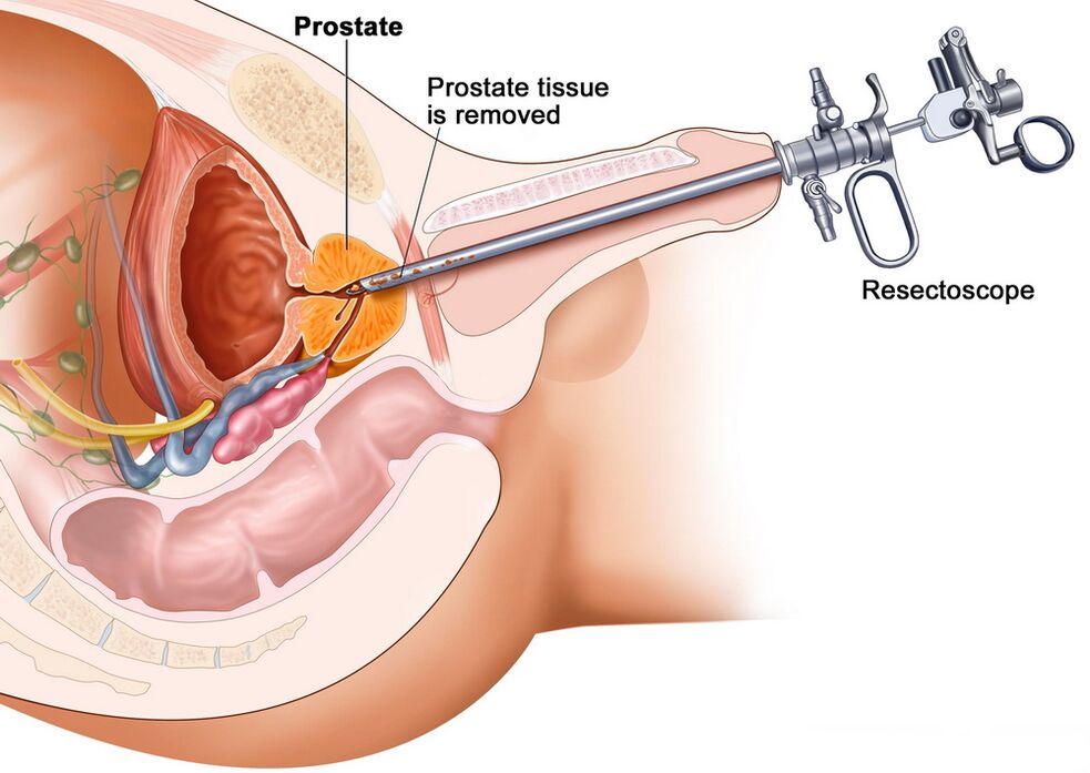
Prostate tissue
During the examination of an enlarged prostate, it is not always possible to understand the nature of the seals and protrusions with the help of a rectal examination and urine and blood tests. It can be a benign pathology (adenoma, prostatitis) or malignant (cancer). Accurate diagnosis helps the microscopic examination of prostate tissue, which is obtained by biopsy.
The procedure is performed as follows: the patient is inserted transrectally with an ultrasound machine sensor, at the end of which there is a gun with a biopsy needle. With a sharp tip, a microscopic part of the gland tissue is cut and given to the laboratory for study. The examination is carried out according to the method of comparing the parameters of the material with the rates from the Gleason table.
With congestive, viral or bacterial prostatitis, the cells of the gland appear reduced in size, the amount of connective tissue in the intercellular space is increased. Atypical cells with altered nuclei will not be seen. If a man has prostate cancer, then the cells of the glands become large and gather in groups, their abnormal modifications are revealed.
Ultrasound, MRI and other methods
To confirm the diagnosis, as well as to determine the stage of development and features of the course of the disease, instrumental studies are carried out. For pathologies of the pelvic organs, the following examination methods are used:
- traditional ultrasound;
- transrectal ultrasound;
- magnetic resonance imaging (MRI);
- CT scan.
These methods allow you to detect the shape, thickness, width, length of the prostate, its mass, structural uniformity, echogenicity, vascularization (vascular pattern). These parameters are needed to determine urological pathologies: ultrasound, CT and MRI show inflammatory, proliferative, oncological diseases of the prostate gland.
Classic ultrasound has the greatest inaccuracy, but this method continues to be used, as it is easy to use and affordable. Transrectal ultrasound is considered the "gold standard" in detecting prostatitis, but prostate cancer is difficult to see this way (especially in the early stages). MRI and CT have the highest accuracy in determining tumors, but these are complex and expensive procedures, so they are performed when other research methods show a high probability of oncology.
Examination at home
The prostate can be examined at home and the main symptoms of urological pathologies can be identified. Of course, this will not be a diagnosis of chronic prostatitis, since it will not be possible to reliably determine the cause of the enlarged gland. But the presence of alarming signs during an independent examination of one's body is a significant reason to immediately contact a urologist.
Like this, without the need to carry out self-diagnosis is not worth it. Indications to be examined at home are:
- Impaired urodynamics (frequent urge to urinate).
- Poor flow, inability to empty the bladder completely.
- Abdominal or groin discomfort (for example, painful urination).
- Decreased sexual desire, weakening of erection.
- Purulent impurities or discoloration of urine in white, brown.
- Spermatorhea or prostorrhea (discharge from the penis).
At home, the examination is done according to the same scheme as in the doctor's office. First, a man must clean the intestines - in 10-12 hours, perform an enema or take laxatives. Take a shower immediately before the procedure. Then lie on your side, bend your knees, insert your index finger into the rectum (first put it on the tip of your finger and smear it with petroleum jelly on top).
Digital rectal examination is performed by observing the posterior wall of the intestine and revealing the adjacent prostate. The gland is easily detected - it feels like a small nut to the touch. Bad symptoms: enlarged prostate, non-circular shape, presence of tuberculosis, pain during probing.These signs signal inflammation or other pathological processes of the prostate gland. When they are identified, you should definitely go to the urologist, as a more accurate diagnosis and treatment plan is required.